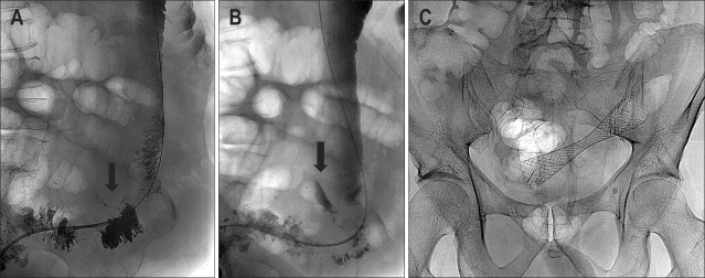
Fig. 9.
(A) Hydrosoluble contrast-agent enema during stent deployment shows limited perforation (arrow). (B) A delayed film shows focalized collection of the contrast agent (arrow). (C) The stent was deployed and the patient was carefully observed for signs of peritonitis. Radiograph showing the stent in an adequate position. The patient underwent elective surgery 12 days later, during which a limited area of inflammation was noted around the perforation.

