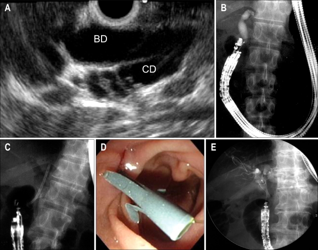
Fig. 2.
Endoscopic ultrasonography-guided choledochoduodenostomy (EUS-CDS). (A) Convex echoendoscope, located in the apex of the duodenal bulb, clearly displayed the extrahepatic bile duct and cystic duct. (B) The echoendoscope was observed in the long/pushing scope position. Cholangiogram obtained by EUS-guided puncture with the tip of the convex transducer directed to the hepatic hilum. (C) Choledochoduodenostomy was accomplished using a plastic stent in the apex of the duodenal bulb. (D) The plastic stent was visible in the first portion of the duodenum. (E) The covered metal stent was also available for EUS-CDS.

