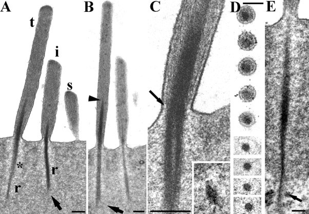
Figure 2.
Structure of the rootlets in rat OHCs. A, Typical example of a hair bundle in a 40 nm vertical ultrathin section from an apical location. The tall (t), intermediate (i), and short (s) stereocilia are visible and rootlets (r) of the tall and intermediate rows; the short rootlet is out of the plane of section. A lighter zone is apparent around the rootlet (asterisk) and splaying is visible at the lower end of the intermediate rootlet (arrow). B, Similar view in a semithin (250 nm) section which has captured the full width and length of the rootlets (as determined from adjacent serial sections). Note the tapering of the rootlet into the shaft, particularly of the tall stereocilium (arrowhead), and the splaying of the end of the tallest stereociliary rootlet in the cuticular plate (arrow). C, The ankle region of a tall stereocilium, showing the filaments in the stereocilium shaft converging onto the rootlet (arrow shows this on one side). Inset, A cross section of a rootlet deep in the cuticular plate showing the crescent shaped profile. D, Serial sections of a single stereocilium through the ankle region. The rootlet is the dense central material. E, Example of a rootlet extending through the lower edge of the cuticular plate (arrow) into the apical cytoplasm, where it splays out. Scale bars: A–C, inset, 200 nm; D, 100 nm; E, 150 nm.

