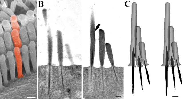
Figure 4.
Serial section reconstruction of representative stereocilia from an apical hair bundle. A, Scanning electron micrograph of a guinea pig OHC bundle to illustrate the concept of a “column” of stereocilia (colored red). B, Two sequential sections of the nine semithin (200 nm) serial sections of a rat OHC used to construct a column of stereocilia as illustrated in A. Note the dense rootlet like material in the upper region of the tall stereocilium displaced toward the edge (arrow). This is discontinuous with the normal rootlet material. C, Stereopair of the reconstructed column. Note how the thickness and length of the rootlet is approximately proportional to the height of the stereocilia in each row. The upper dense material has been omitted for clarity. Scale bars, 200 nm.

