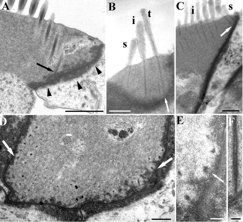
Figure 7.

A, Vertical ultrathin section of an apical rat OHC showing a row of rootlets sectioned obliquely, one of which joins the lower end of the lateral junctional complex (arrow). Note the lip around the cell apex (arrowheads). B, Semithin section of a basal rat OHC showing contact between the rootlet of the tall stereocilia (t) and the lateral wall (arrow). Intermediate stereocilia (i) and short stereocilia (s) rootlets also closely approach the wall. C, Another basal rat OHC where short stereociliary rootlets contact the lateral wall (arrow). Intermediate stereocilia are also visible (i). D, Horizontal section of an apical rat hair cell showing the rootlets on either end of the bundle closely in contact with the lateral wall (arrows). E, Detail of a serial section to that shown in D. Rootlets approach the junctional complex and some appear to have filaments extending into it (arrow). F, Vertical section of a rootlet contacting the lateral wall in a rat hair cell. The dense material appears to be contiguous. Scale bars: A, 1 μm; B–D, 0.5 μm; E, F, 100 nm.
