
Figure 8.
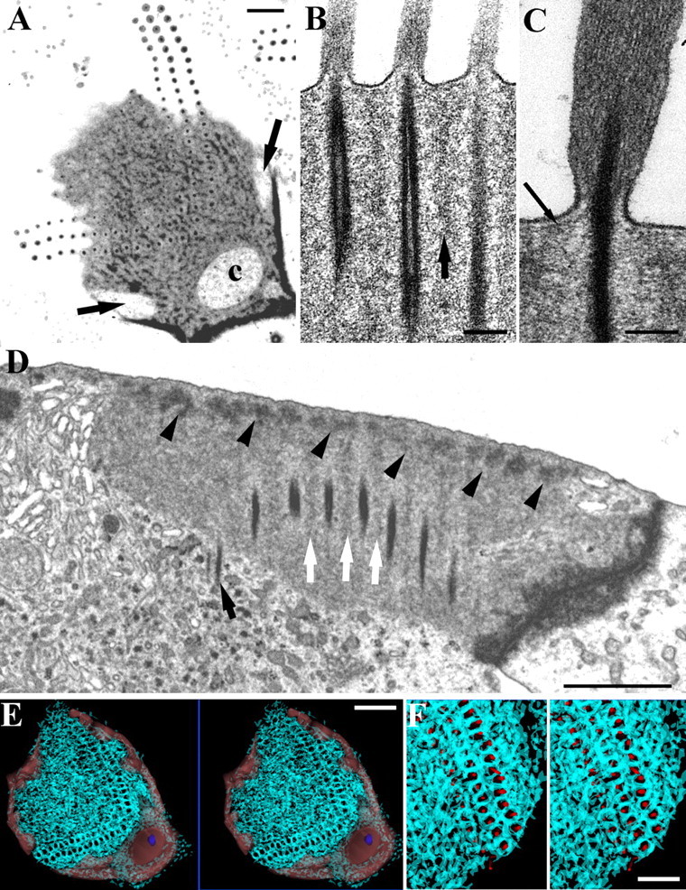
The structure of the meshwork in the cuticular plate. A, Horizontal section of a guinea pig OHC. The dense material forms a mesh around the rootlets extending over the superficial aspect of the plate. Channels in the periphery of the cuticular plate (arrows) and at the apex of the W (c) are visible. B, Vertical section of a rat OHC, showing vertically orientated parallel actin filaments between the rootlets (arrow). C, A subapical dense layer extends over the cuticular plate upper surface and contacts the rootlet where it enters the plate (arrow); rat OHC. D, Vertical section of a rat OHC showing dense material in the apical meshwork along the upper edge of the plate (arrowheads). Note lighter material extending down from the mesh (white arrows) and one rootlet penetrating into the cytoplasm below the cuticular plate (black arrow). E, F, Two sets of stereopairs at low and intermediate magnification of the meshwork reconstructed from serial sections of a guinea pig OHC (cyan traces). The channels are indicated in light red in E and the basal body (normally found in the large channel on the strial side of the bundle) is shown in blue. Reconstruction shows that the meshwork around the rootlets extends in 3D so that each is ensheathed by the dense/filamentous material of the meshwork. F, Rootlets are shown as red cylinders sitting in their sheaths. Scale bars: A, D, F, 1 μm; B, C, 100 nm; E, 2 μm.