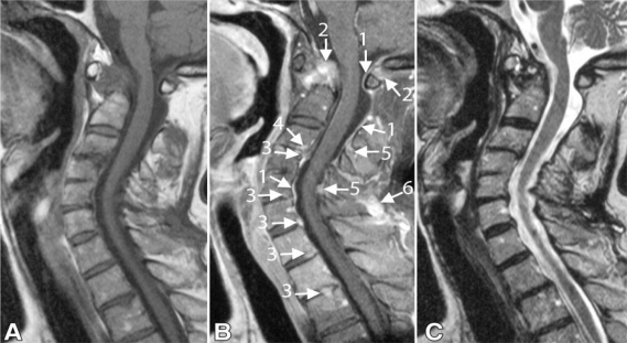
Figure 2.
Sagittal fast spin-echo MRI of the cervical spine in an RA patient (a 51-year-old woman who had suffered RA for 31 years) with superficial and deep enhancement. A) (T1-weighted) and C- (T2-weighted) images show; stenoses at levels C1–2 and C3–4, mostly caused by pannus and subluxation at level C1–2 and by discopathy and ligamentum flavum hypertrophy. B) (gadolinium-enhanced T1-weighted image) shows; superficial enhancement lining the cerebrospinal fluid (arrow 1) and enhancement involving deeper structures. Deep enhancing tissue is recognized as bone and pannus tissue on C1–2 (arrow 2), as a disc on anterior levels (arrow 3), level C3–4 (arrow 4). Ligamentum flavum and interspinal ligaments are enhanced at the posterior levels (arrow 5). Deep enhancement coincides mostly with narrowing of the spinal canal at these levels. Note enhancement of the ligamentum nuchae (arrow 6).

