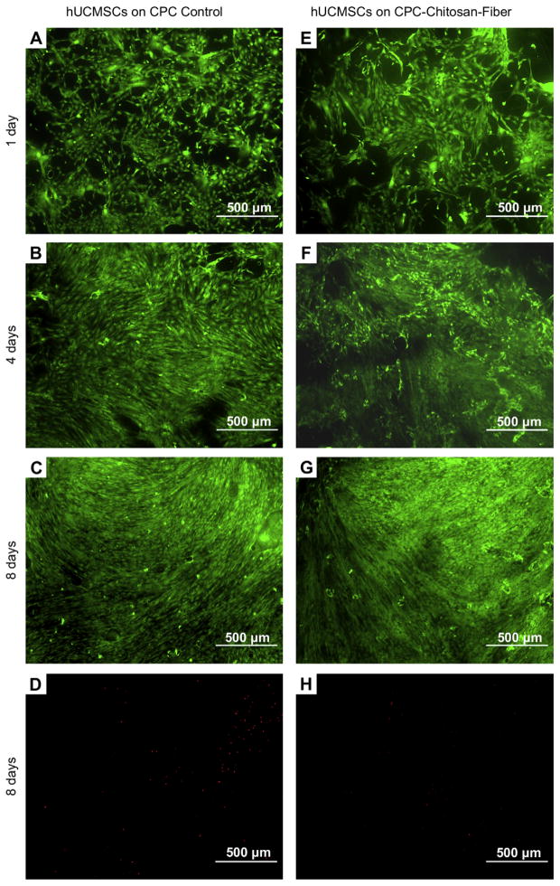
Fig. 4.
Live/dead staining of hUCMSCs cultured on CPC control and CPC–chitosan–fiber for 1 day, 4 days, and 8 days. Live cells, stained green, were numerous on both materials. Dead cells, stained red, were few on both materials. Three randomly-chosen fields of view were photographed from each disk. A total of five disks yielded 15 photos per material at each time period. Representative photos are shown here.

