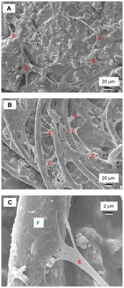
Fig. 6.
SEM of hUCMSC attachment on: (A) CPC control, and (B) CPC–chitosan–fiber scaffold. Cells are designated as “C”, which anchored to CPC in (A), and to the fibers in the scaffold in (B). Cells developed long, cytoplasmic extensions “E”, shown in (C) at a higher magnification, attaching firmly to the fiber in the CPC–chitosan–fiber scaffold.

