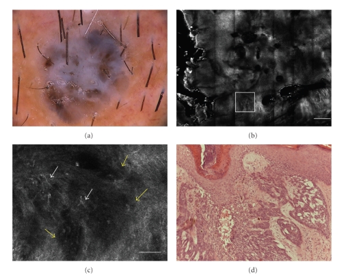
Figure 1.
(a) Dermoscopic image of nodular pigmented lesion showing a blue gray veil structureless brown areas and dark brown dots in the center (original magnification 30×). (b) RCM image (4 × 4 mm) at dermoepidermal level, showing a general cauliflower architecture highly specific for BCC diagnosis (scale bar: 500 μm). (c) RCM image (0.5 × 0.5 mm, white frame), showing dark silhouettes (yellow arrows) outlined by dark spaces. In the inner portion of them, bright filaments were present (white arrows) (scale bar: 50 μm). (d) Histology showing basaloid nodules in the dermis corresponding to hypopigmented dark silhouettes. Within and outside the tumor nests some melanin was present (H&E section; original magnification ×100).

