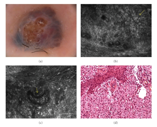
Figure 4.
(a) Dermoscopy showed structureless areas with blue hue and a globular pattern. Ulceration was present in the center of the nodule (original magnification 30×). (b) RCM image (0.5 × 0.5 mm) at dermal level, showing pleomorphic small cells (yellow arrow) arranged to form a cerebriform nests delimitated by fibrous septa (scale bar: 50 μm). (c) RCM image (0.5 × 0.5 mm) at dermal level, showing an enlarged new-born vessel with a prominent blood flow (yellow arrow) (scale bar: 50 μm). (d) Histology showing a solid proliferation of atypical melanocytes in which new-born vessels can be recognized (H&E section; original magnification ×100).

