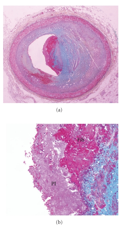
Figure 4.
Acute erosion, with early organization, Movat pentachrome. (a) Low magnification of the left anterior descending coronary artery with severe luminal narrowing and nonocclusive thrombus. The underlying plaque is rich in smooth muscle cells, without significant lipid and no core formation. (b) Higher magnification demonstrating a single layer of fibrin in the proteoglycan-rich cap (right). The center of the photomicrograph demonstrates layering of the thrombus, with platelets (Pl) adjacent to the lumen overlying fibrin (Fib).

