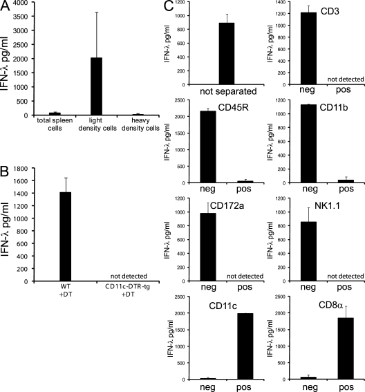
Figure 4.
The IFN-λ production to poly IC injection in vivo separates with CD45R−/CD11c+/CD8α+ splenocytes. 1.5–2 h after i.v. injection of poly IC, spleens were harvested and processed. Cell-free supernatants were analyzed for IFN-λ after in vitro culture for 18 h. (A) 5 × 10⁶ cells/ml of total spleen cells or cells separated by density centrifugation into light density cells or heavy density cells. (B) 25 × 106 cells/ml of total spleen cells of WT or CD11c-DTR-tg mice treated 2 d before with DT. (C) Total spleen cells before separation or after magnetic bead separation into the denoted populations. The initial cell number of splenocytes added onto the column was 20 × 106. Without further counting, each fraction was distributed into 2 wells with 200 µl of medium/well. Bars represent the mean ± SD of two independent experiments (A and C) or one experiment (B) using two mice per experiment.

