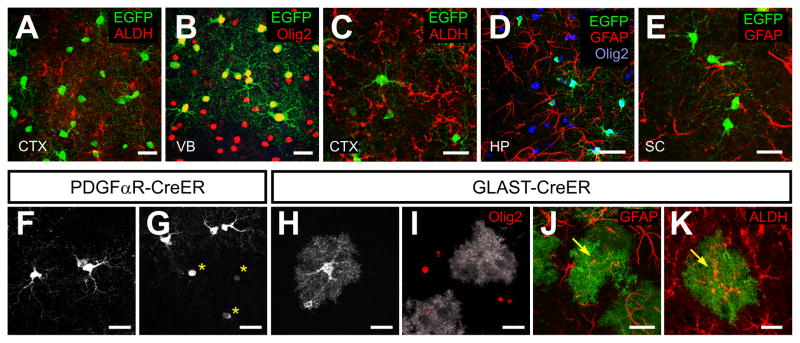
Figure 5. NG2+ cells in the postnatal brain do not generate astrocytes.
(A – E) Confocal images showing immunoreactivity to EGFP, ALDH, Olig2 and GFAP in the dorsal cortex (CTX; A, C), ventral forebrain (VB; B), hippocampus (HP; D), and spinal cord (SC; E) in PDGFαR-CreER;Z/EG mice at P4+96 (A and B) or P30+120 (C – G).
(F – K) Comparison of the morphologies of EGFP+ cells from PDGFαR-CreER;Z/EG mice at P30+120 (F, G), GLAST-CreER;Z/EG at P11+10 (H) or GLAST-CreER;ROSA26-mGFP mice at P15+21(I – K). Confocal images of EGFP+ cells from cortex (F – I), hippocampus (J) or ventral forebrain (K). Yellow asterisks highlight mature OLs, in which cytosolic EGFP is visible only within the soma (G). Yellow arrows in J and K show EGFP+ astrocytes that were GFAP+ or ALDH+. Scale bars = 20 μm.

