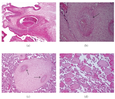
Figure 1.
Characteristic histological findings of placental vascular obstructive lesions (all images colored by hematoxylin and eosin; magnification 40×). (a) Occlusive thrombus (arrow) in a dilated chorionic vein. (b) Hemorrhagic endovasculitis (HEV): extravasated blood cells (arrow) around a vessel in a stem villus. (c) Muscular hypertrophy and old occlusions (arrows) in stem vessels (obliterative endarteritis) (OE). (d) Avascular villi demonstrating hyaline villous stroma devoid of vessels.

