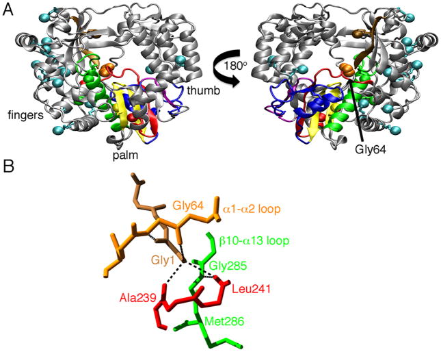
Figure 1.
Structure of PV 3Dpol (PDB 1RA6). A. The locations of the terminal methyl groups of the methionines are indicated as colored spheres. The conserved structural motifs in the palm subdomain are colored as follows: motif A, red; motif B, green; motif C, yellow; motif D, blue; and motif E, purple. Gly64 is colored orange, and the N-terminal β-strand is colored brown. B. Hydrogen bond interactions involving Gly64, Gly1 on the N-terminal β-strand. Ala239 and Leu241 on motif A, and Gly285 on the β10-α13 loop. Motifs are colored as in A. Hydrogen bonds are indicated as black lines.

