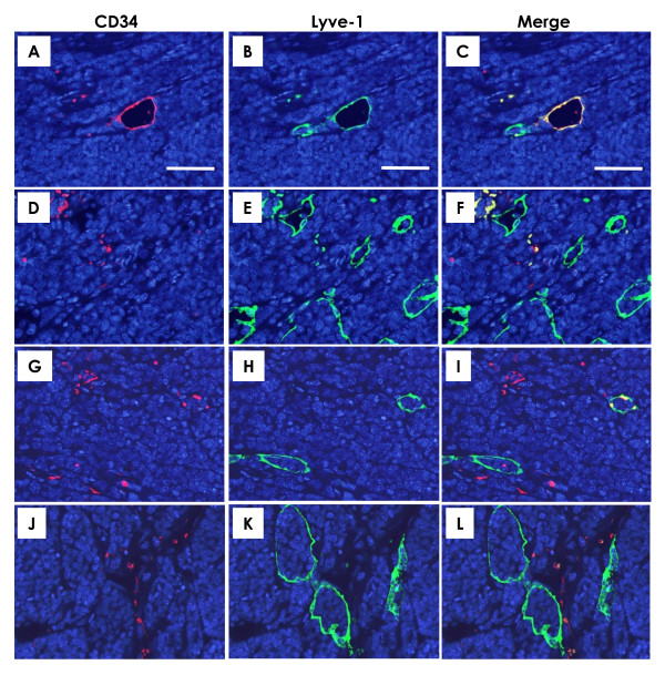
Figure 4.
Immunofluorescence for blood and lymphatic vessels in mammary carcinomas. Double immunohistochemical staining with CD34 for blood microvessels (red) and Lyve-1 for lymphatic (green) microvessels and their merged images in mammary tumors. As can be seen in their merged image (C), some microvessels showed both expressions of CD34 (A) and Lyve-1 (B). Such microvessels having CD34+/Lyve-1+ characteristics were excluded from quantitation. The number of lymphatic microvessels was lower in the pesVEGFR-2 (G-I, merge in I) and pEndo (J-L, merge in L) groups than in the pVec group (D-F, merge in F). A-L, scale bar = 50 μm.

