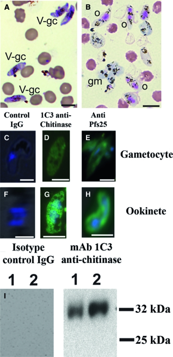
Figure 4.

Light and fluorescence microscopy of in vitro cultured P. falciparum sexual stage parasites. (A) Leukostat-stained thin smears of unpurified gametocyte culture contained stage V gametocytes (V-gc). (B) Leukostat-stained thin smears of in vitro cultured P. falciparum sexual stage parasites. Ookinetes (o) were identified by the presence of one to two large eosinophilic nuclei and lack of a surrounding erythrocyte membrane. Round forms include macrogametes and zygotes. Self-adherent macrogametes (gm) were slightly basophilic and often found in clusters. Non-adherent macrogametes were not definitively distinguished from zygotes and retort ookinetes. Hemozoins appeared as dark brown pigment crystals. (C–H) Gametocytes from gametocyte cultures and ookinetes from sexual stage cultures were probed with antibodies to chitinase and Pfs25. DAPI (blue) was used to stain nuclear material; gametocytes contained one nucleus, whereas ookinetes contained one to two nuclei. (C) Gametocytes and (F) ookinetes probed with IgG isotype control antibody showed no reaction. (D) Gametocytes and (G) ookinetes probed with 1C3 monoclonal antibody against chitinase45 (green) showed a diffuse, intracellular staining pattern in both gametocytes and ookinetes. (E) Gametocytes and (H) ookinetes probed with antibody against Pfs2558 (green) showed an intracellular pattern in gametocytes and both intracellular and surface staining patterns with ookinetes. (I) Chitinase detected in gametocytes and a 72-hour cultured mixed zygotes and ookinetes sample by Western immunoblot. Equal numbers of cells gametocytes (lane 1) and untransformed gametes plus zygotes plus ookinetes (lane 2) were lysed in SDS, separated by SDS-PAGE, and probed with antibody against the P. falciparum chitinase PfCHT1 using monoclonal antibody 1C3.45 IgG isotype control antibody did not produce a band in either sample. Antibody against PfCHT1 recognized an approximately 32-kDa band in both gametocyte and zygote plus ookinete samples and quantitatively more after 72 hours of cultures than in gametocytes.
