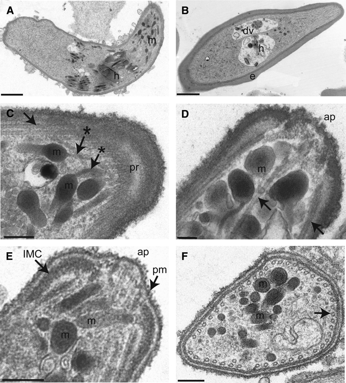
Figure 5.

Ultrastructural features of the P. falciparum ookinete. (A) Sagittal section of an ookinete shows micronemes (m), seen as electron-dense round or cigar-shaped organelles, and a hemazoin crystal (h). (Scale bar: 1 μm.) (B) For comparison, a gametocyte within an infected erythrocyte (e) is seen with a discrete digestive vacuole (dv) with hemozoin (h). (Scale bar: 1 μm.) (C) Tangential section of the apical end of an ookinete showed microtubules (→), which converge at the polar ring (pr). This section showed narrow ducts of micronemes (m) that appear to track to the apical end (*→). (Scale bar: 200 nm.) (D) Transverse, midline section of an ookinete apical end showed the apical pore (ap), micronemes (m), and microtubules (→). (Scale bar: 200 nm.) (E) Transverse section of an ookinete showed micronemes (m) as well as the apical pore (ap) located between two electron-dense regions representing the inner membrane complex (IMC) underlying the plasma membrane (pm). (Scale bar: 200 nm.) (F) Transverse cross-section near the apical end of an ookinete shows micronemes (m) as well as microtubules (→) that circumferentially line the subpellicular space of the parasite. (Scale bar: 200 nm.)
