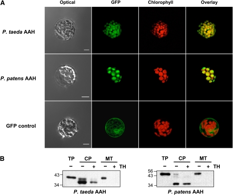
Figure 6.
Evidence That the P. taeda and P. patens AAH Proteins Are Chloroplast Targeted.
(A) Transient expression in Arabidopsis mesophyll protoplasts of GFP fused to the C terminus of P. taeda AAH (top panels), P. patens AAH (middle panels), or GFP alone (bottom panels). GFP (green pseudocolor) and chlorophyll (red pseudocolor) fluorescence were observed by confocal microscopy. Bars = 10 μm.
(B) Protein import into isolated pea chloroplasts and mitochondria. Full-length P. taeda and P. patens AAH sequences were translated in vitro in the presence of [3H]Leu. The translation products were incubated for 15 (P. taeda) or 10 (P. patens) min in the light with chloroplasts (CP) or mitochondria (MT), which were then repurified on an 8% (v/v) Percoll gradient, without or with prior thermolysin (TH) treatment to remove adsorbed proteins. Proteins were separated by SDS-PAGE and visualized by fluorography. Samples were loaded on the basis of equal chlorophyll or mitochondrial protein content next to aliquots of the respective translation products (TP). The positions of molecular mass standards (kD) are indicated.

