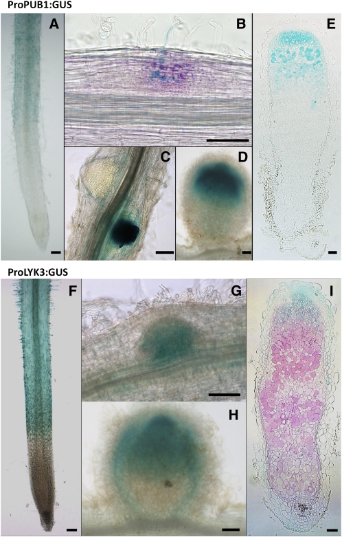
Figure 5.
Histochemical Localization of ProPUB1:GUS and ProLYK3:GUS Expression during Nodulation of M. truncatula.
PUB1 expression pattern was determined using a 3-kb promoter region of the gene cloned in front of the GUS coding sequence. LYK3 expression pattern was determined using the 2.6-kb promoter region of LYK3 cloned in front of the GUS-GFP fusion coding sequence. M. truncatula roots transformed by A. rhizogenes were used for ProPUB1:GUS or ProLYK3:GUS detection during nodulation using either X-Gluc (blue in [A] and [C] to [I]) or Magenta-Gluc (magenta in [B]). Rhizobia were detected by β-galactosidase (LacZ) activity using X-Gal (blue in [B]) or Magenta-Gal (magenta in [I]). Bars = 100 μm.
(A) ProPUB1:GUS is weakly expressed in noninoculated roots, including in the root hair development zone.
(B) After inoculation, ProPUB1:GUS is expressed predominantly in the nodule primordium and in cells associated with infection.
(C) ProPUB1:GUS is expressed strongly in nodule compared with root primordia.
(D) ProPUB1:GUS is expressed in the apical region of young, developing nodules.
(E) In mature nodules, ProPUB1:GUS is expressed in the apical region, including the preinfection and infection zones and the early nitrogen-fixing zone.
(F) ProLYK3:GUS is expressed in the developing root hair zone of noninoculated roots, including the epidermal cells.
(G) ProLYK3:GUS is expressed in the nodule primordium.
(H) In young nodules ProLYK3:GUS is expressed predominantly in the apical region of the nodule and to some extent in the vascular tissue.
(I) In mature nodules, ProLYK3:GUS expression is predominantly in the apical region, including the preinfection and infection zones.

