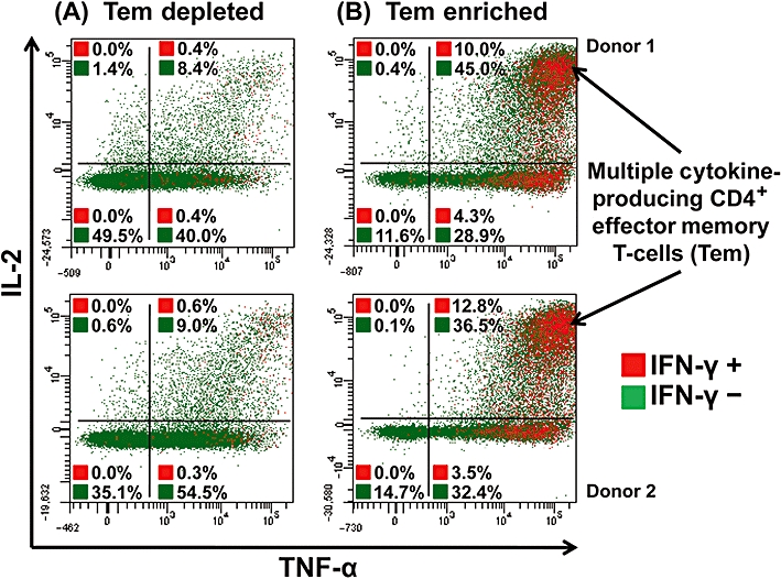
Figure 5.

Intracellular cytokine staining of human CD4+ effector memory T-cell (Tem)-depleted (A) and -enriched (B) PBMCs for TNF-α, IL-2 and IFN-γ, following stimulation with 1 µg per well immobilized TGN1412. Tem-depleted (left panels) and Tem-enriched (right panels) PBMCs were surface stained for T-cell markers and intracellular cytokines; analysis shown is gated on CD4+ T-cells. Cells in the right-hand quadrants are positive for TNF-α, cells in the upper quadrants are positive for IL-2, cells shown in red are positive for IFN-γ and cells shown in green are negative for IFN-γ. Cells in the upper right corner in red are positive for TNF-α, IL-2 and IFN-γ. The frequency of cytokine positive cells is expressed as a percentage of CD4+ T-cells. Data from two representative donors are shown (similar data were obtained with four additional donors from two independent experiments).
