
Figure 6.
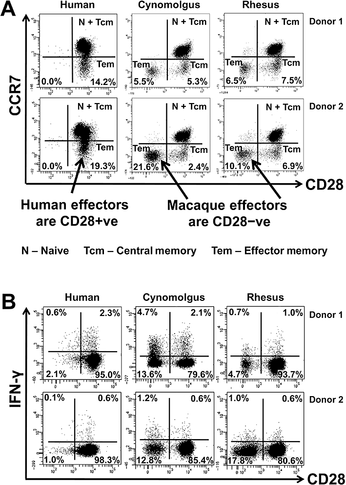
Comparative immunophenotyping of human, cynomolgus and rhesus macaque CD4+ T-cells. All analysis shown is gated on CD4+ T-cells. (A) Analysis of memory markers using CD28 and CCR7 staining. Human, cynomolgus and rhesus macaque naive CD4+ T-cells (CCR7+CD28+) and central memory T-cells (CCR7+CD28+), macaque transitional memory T-cells (CCR7-CD28+) and human effector memory CD4+ T-cells (CCR7-CD28+) all express CD28. In contrast, both cynomolgus and rhesus effector memory T-cells (CCR7-CD28-) do not express CD28. Data from two representative donors are shown (similar data were obtained with two additional rhesus, two cynomolgus and six human donors from two independent experiments). (B) Intracellular IFN-γ staining versus CD28 expression on CD4+ T-cells following PMA and ionomycin stimulation for 4 h. Human CD4+ T-cells constitutively express CD28, so IFN-γ staining stems only from the CD28+ population. Macaque CD4+ T-cells can be subdivided into CD28+ (naive, central and transitional memory T-cell) and CD28- (effector memory T-cell) populations, both of which produce IFN-γ following PMA and ionomycin stimulation for 4 h. A greater fraction of macaque CD4+CD28- T-cells produce IFN-γ compared to CD4+CD28+ T-cells following mitogen stimulation (similar data were obtained with two additional rhesus, two cynomolgus and two human donors).