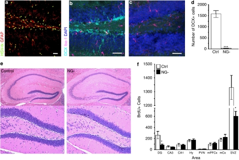
Figure 1.
(a) Immunohistochemistry against HSV-tk and GFAP in the hippocampal dentate gyrus (DG) of adult hGFAPtk mice reveals HSV-tk expression in GFAP+ cells. (b–d) The DG of control mice contains many doublecortin (DCX) positive neuroblasts (aqua) while these cells are virtually abolished (F8,8=428.2, Student's t-test P<0.0001) in hGFAPtk mice treated with VGCV for 3 weeks (NG-, 26.67±7.688, n=9, gray bar; Controls, 1577±159.1, n=9, white bar) . Staining for Iba1 (purple) reveals no increase in activated microglia. Scale bars=30 μm. (e) Hematoxylin and Eosin (H&E) staining shows no indication of inflammation in the hippocampus of NG- mice treated with VGCV for >8 weeks. (f) Analysis of cell proliferation and short-term survival in the adult CNS of Ctrl and NG- mice. After >8 weeks of VGCV treatment mice were injected twice daily for 5 days with BrdU (200 mg kg–1 body weight) and perfused 24 h after the last injection. BrdU+ cells were counted in the hippocampal DG and Cornu Ammonis regions (CA3 and CA1), in the hypothalamus (Hy) including the paraventricular nucleus (PVN), as well as in medio-prefrontal cortical regions (mPFCx), motor cortex (mCx) and the subventricular region of the lateral ventricles (SVZ). A decrease in BrdU+ cells was only detected in the DG (F3,5=12.27, Student's t-test P=0.0155) and the SVZ (F3,5=1.691, Student's t-test P<0.001). (Control, n=4, white bars, NG- n=6, black bars) Note that NG- mice still show a high rate of cell proliferation in the DG and SVZ. This is most likely due to the generally high proliferative capacity in these regions including cell types of non-neuronal lineages that do not undergo differentiation into DCX+ positive neuroblasts such as microglial cells, endothelial cells as well as oligodendrocyte progenitor cells.

