Figure 3.
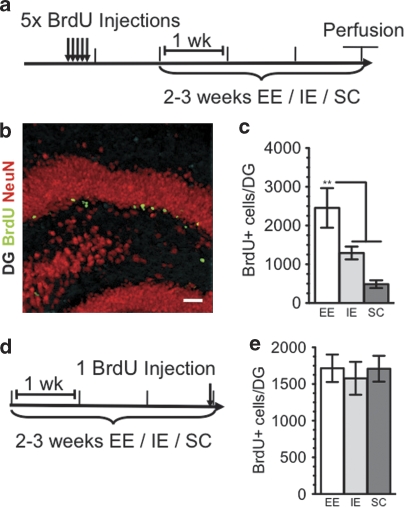
(a) Timeline shows BrdU injections for studying survival of newborn neurons under different housing conditions. Animals were injected with BrdU (200 mg kg–1) 1 × per day for 5 days, 1 week before being placed in either an enriched environment (EE) or an impoverished environment (IE) for 3 weeks before perfusion. In the social conflict (SC) condition, animals were injected 1 × per day for 5 days, 2 weeks before being placed in SC for 2 weeks before perfusion. (b) Newborn neurons were analyzed using double labeling for BrdU (green) and the neuronal marker, NeuN (red). Almost all BrdU-labeled cells also expressed NeuN. (c) Animals living in EE displayed significantly increased survival of newborn neurons as compared with either animals in IE or SC (EE, 2838±562.8, n=6, white bars; IE, 1291±164.8, n=5, gray bars; SC, 572.0±45.18, n=6, dark gray bars; F2,14=10.98, P=0.0014; Tukey-Kramer post hoc test for EE vs IE P<0.05, for EE vs SC P<0.01). (d) Timeline shows BrdU injections for studying proliferation of newborn neurons under differential housing conditions. Animals were given a single injection of BrdU 3 h before being euthanized after having lived in either EE, IE or SC housing conditions for 2–3 weeks (EE, 1715±530.5, n=8, white bars; IE, 1578±634.2, n=8, gray bars; SC, 1709.0±499.2, n=8, dark gray bars).

