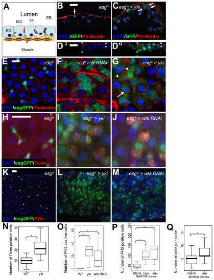
Fig. 1.
Loss of Hpo signalling promotes ISC proliferation. (A) The adult midgut. (B,C) Orthogonal cryosections of the adult midgut epithelium showing that esgts-driven expression of Yki-GFP (C) leads to increased nuclear density and the number of basally located esg+ cells (arrows) compared with control (esgts>GFP) (B). Nuclei are stained with DAPI in all panels (blue), esg+ cells are marked by GFP (green) and phalloidin staining is red. (D,D′) Orthogonal section of a 2-week-old MARCM hpo clone shows increased epithelial thickness (arrows) compared with surrounding GFP-negative control tissue. Progenitor cells are marked by armadillo (β-catenin) staining (red). Confocal micrographs of adult posterior midguts. (E-G) esgts-driven expression of Yki (G) or Notch-RNAi (F) in ISCs and EBs induces an increase in esg+ (green) cell number compared with control (E). Yki overexpression induces the appearance of esg+ cells with large nuclei (arrow), but smaller nuclei remain (arrowheads) (G, compare with E and F). Phalloidin is in red. (H-J) esgts-driven expression of Yki (I) or wts-RNAi (J) induces increased numbers of Dl+ cells compared with control (H). esg+ cells are marked by GFP (green) and Dl is in red. (K-M) esgts-driven expression of Yki (L) or wts-RNAi (M) increases the number of PH3+ cells compared with control (K). esg+ cells are marked by GFP (green) and PH3 is in red. Scale bars: 10 μm in E-G; 20 μm in B-D′,H-M. (N) Quantification of Dl+ cells. esgts-driven expression of Yki significantly increases the total number of Dl+ cells in adult midguts compared with control. P<0.001, n>10. (O) Quantification of PH3+ cells. esgts-driven expression of Yki or wts-RNAi significantly increases the total number of PH3+ cells in adult midguts compared with control. In both cases, P<0.0001, n>15. (P) hpo42-47 or savshrp1 mutant MARCM clones increased mitotic rates (PH3+ cells/gut) along the entire midgut compared with control clones. P<0.0001, n=12. (Q) Increased cell number in 7-day-old savshrp1 mutant MARCM clones (n=46) compared with neutral MARCM clones (n=74). P<0.0001.

