Fig. 8.
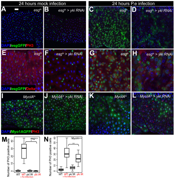
Yki is required for the midgut regenerative response to bacterial infection. (A-H) P.e infection induces a proliferative response with increased numbers of esg+ cells, mitoses, Dl+ ISC-like cells and midgut size (A,C,E,G). esgts-driven expression of yki-RNAi (B,F) causes a reduction in the midgut regenerative response to infection (D,H). (I-L) MyoIAts-driven expression of yki-RNAi (J) partially prevents the midgut regenerative response to stress (L). Nuclei are stained with DAPI (blue); esg+ cells (A-H) and ECs (I-L) are marked by GFP (green); PH3 (A-D, I-L) and Dl (E-H) are red. (M,N) Quantification of PH3+ cells upon bacterial infection. esgts-driven expression of a yki-RNAi construct prevents the regenerative response seen in wild-type midguts upon bacterial infection (M) (*P<0.001, n>10). MyoIAts-driven expression of yki-RNAi partially rescues the midgut regenerative response following bacterial infection (N) (**P=0.018, n>14). Scale bar: 20 μm.

