Fig. 4.
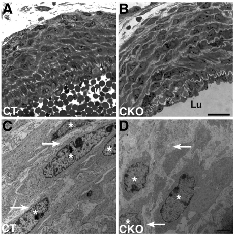
Ultrastructural changes in the ductus arteriosus wall of Jag1-SMcko mouse embryos. (A-D) The tunica media of E18.5 control littermate ductus arteriosus (A) is more compactly organized than that of the Jag1-SMcko ductus arteriosus (B), particularly in layers distal to the vessel lumen (Lu). The ductus arteriosus medial wall of the control littermate (A) is composed of vascular smooth muscle cells with spindle-shaped nuclei (asterisks in high-magnification view C) forming compact, well-organized layers throughout the width of the vessel wall. These cells are surrounded by thin layers of extracellular matrix (arrows in C). The Jag1-SMcko ductus arteriosus is composed of more loosely organized cells (B), with irregularly shaped nuclei that are surrounded by thicker layers of extracellular matrix (arrows in high-magnification view D) than in the control (C). Asterisks mark smooth muscle nuclei. Scale bars: 20 μm in A,B; 2 μm in C,D.

