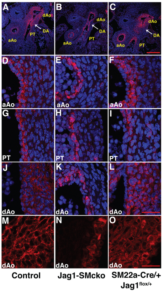
Fig. 6.
JAG1 protein is highly expressed in the ductus arteriosus and descending aorta and is required for its own expression. Transverse sections of E13.5 mouse embryos through the level at which the ductus arteriosus (DA) connects to the descending aorta (dAo). Sections are immunostained with JAG1 antibody (red); A-L are also counterstained with DAPI (blue) to highlight nuclei. (A-C) Low-magnification views showing the JAG1 expression pattern in the ductus arteriosus and descending aorta, ascending aorta (aAo) and pulmonary trunk (PT). (D-L) High-magnification views of the medial walls of the ascending aorta (D-F), pulmonary trunk (G-I) and descending aorta (J-L). (M-O) JAG1 immunofluorescent staining reveals the superimposition of the wild-type JAG1 and mutant JAG1 expression patterns throughout the entire medial wall in the descending aorta of SM22a-Cre/+; Jag1flox/+ embryos (O). Scale bars: 200 μm in A-C; 40 μm in D-L; 25 μm in M-O.

