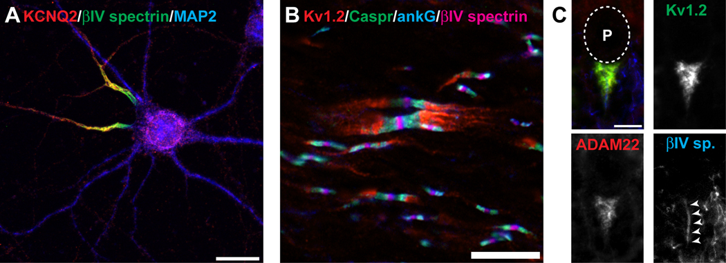
Figure 1. Kv channels are clustered at a many different axonal locations.
A, KCNQ2 K+ channels (red) are clustered at axon initial segments where they colocalize with the AIS-restricted cytoskeletal scaffolding protein βIV spectrin (gree). The microtubule associated protein 2 (MAP2) defines the somatodendritic domain. B, Kv1 channels (red) are clustered at juxtaparanodes beneath the myelin sheath and on each side of nodes of Ranvier. Kv1 channels are excluded from paranodal regions labeled by Caspr (green), and nodes of Ranvier labeled by ankG (blue) and βIV spectrin (magenta). C, Basket cell terminals in the cerebellum are highly enriched in Kv1.2 (green) and ADAM22 (red), and envelope the AIS (labeled by βIV spectrin, blue). The location of the Purkinje neuron cell body (P) is indicated by the dotted line. Scale bars: A, 20 µm; B, C, 10 µm.

