
Figure 3.
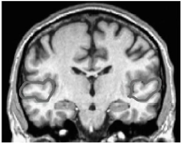
Manually tracing the outer boundaries of the superior temporal gyrus. The superior temporal gyrus was identified bilaterally in reference to standard brain atlases. Each coronal slice containing the STG was manually traced along the gyral boundaries. An automated algorithm was used to multiply cross sectional area by slice thickness to obtain volumetric measurements.