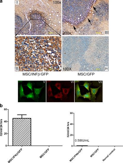
Fig. 1.
MSC/IFNβ/GFP cells home to 4 T1 breast tumors and express high levels of IFNβ. 1 × 10⁶ MSC/IFN-/GFP and MSC/GFP cells were injected into 4 T1 tumor established mice through tail vein. Tissues were collected in 3 days after MSC/IFN/GFP and MSC/GFP cell administration and slides sections were prepared. Immunohistochemical staining with anti-mouse IFN-β antibody in Fig. 1 I-IV indicates that high levels of IFNβ were expressed by MSC especially around the border of the tumor and stromal tissue. Magnifications of the pictures are as indicated. Mice injected with MSC/GFP served as controls, Fig. 1 IV, absence of IFN-β positivity after injection of MSC/GFP cells. Staining attached MSC/IFN-β/GFP cells on slides were probed with anti mouse IFN-β antibody, the secondary antibody is conjugated Rhodamine (Abcam, Cambridge, MA), Fig. 1 VI; or without anti IFN-β antibody, Fig. 1 V. Fig. 1 VII is the merging result for Fig. 1 V and VI. b left, high levels (45 IU/ml) of IFNβ in MSC/IFN-β/GFP culture medium determined by ELISA b right, low levels of IFN-β (0.5-1 IU/ml) in mouse serum collected 3 days after MSC/IFN-β/GFP injection (n = 3)

