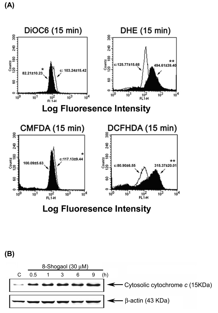
Figure 3. Induction of mitochondrial dysfunction, reactive oxygen species (ROS) generation, GSH depletion, and cytochrome c release in [8]-shogaol-induced apoptosis.
(A) HL-60 cells were treated with 30 µM [8]-shogaol for indicated times and were then incubated with 3, 3'-dihexyloxacarbocyanine (40 nM), DCFH-DA (20 µM), DHE (20 µM), CMFDA (20 µM) respectively and analyzed by flow cytometry. Data are presented as log fluorescence intensity. C: control. (B) Cells were treated with 30 µM [8]-shogaol for 15 min. Subcellular fractions were prepared and cytosolic cytochrome c was analyzed by Western blotting as described in the Material and Methods section. These experiments were performed at least three times, and a representative experiment is presented. *P < 0.05 and **P < 0.01 indicate statistically significant difference from control.

