Figure 1.
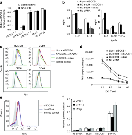
DC3-9dR-mediated SOCS-1 silencing enhances cytokine production and stimulatory capacity of DCs. (a) MDDCs were transfected with three different siRNAs (siSOCS1-1, -2, and -3) targeting different regions of SOCS-1 mRNA using lipofectamine 2,000 or DC3-9dR peptide and stimulated with LPS (100 ng/ml). Twenty-four hours later, SOCS-1 mRNA levels were measured by qRT-PCR. MDDCs treated with an irrelevant siRNA (siLuci) and mock-treated served as controls. (b,c) SOCS1-silenced MDDCs were stimulated with LPS; after 24 hours DC-specific cytokines were quantified from supernatants using a (b) multiplex human cytokine immunoassay (Millipore) and analyzed for costimulatory and MHC molecules by (c) flow cytometry. (d) MDDCs transfected with siSOCS-1 complexed with either lipofectamine or DC3-9dR (1:10 ratio) were cocultured with allogeneic T cells at the indicated ratios for 5 days and proliferation was assayed by 3H thymidine incorporation during the last 16 hours of the assay. (e,f) MDDCs were treated with siLuci or siSOCS-1, and 6 hours later expression of TLR3 was tested by flow cytometry (e) and the expression of the indicated IFN-responsive gene relative to β-actin was analyzed by qRT-PCR (f). Poly (I:C) was used as a positive control. DC, dendritic cell; IFN, interferon; LPS, lipopolysaccharide; MDDC, myeloid-derived DC; MHC, major histocompatibility complex; qRT-PCR, quantitative real-time PCR; siRNA, small interfering RNA; SOCS-1, suppressor of cytokine signaling-1.

