Figure 1.
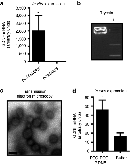
PEG-POD forms GDNF-expressing nanoparticles. (a) A plasmid (pCAGGDNF) containing an expression cassette for rat GDNF was shown to express GDNF mRNA in vitro. Both conditions, n = 3. Mean ± SEM. (b) PEG-POD compaction of pCAGGDNF prevents electrophoretic migration of the plasmid, which can be relieved by trypsin-mediated digestion of the protein. (c) PEG-POD compacted pCAGGDNF (PEG-POD~GDNF) was examined by transmission electron microscopy (TEM) and found to form discrete spherical particles, similar to those previously described. Analysis of the TEM images showed a mean particle diameter of 175.9 ± 28.6 nm. Bar = 200 nm. (d) Injection of PEG-POD compacted pCAGGDNF into the subretinal space results in a detectable level of rat GDNF mRNA expression (P < 0.05). Both conditions, n = 9. Mean ± SEM. GDNF, glial cell line–derived neurotrophic factor; PEG, polyethylene glycol; POD, peptide for ocular delivery.

