Figure 2.
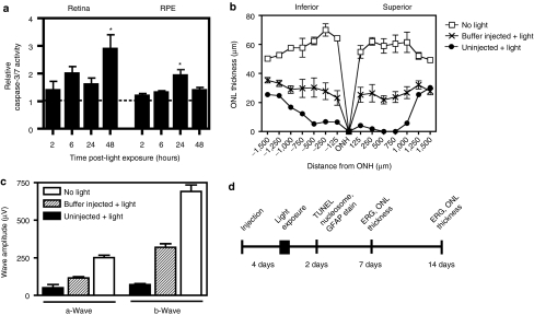
Bright blue light induces caspase-3/7 activation and retinal degeneration that is modulated by subretinal injection. (a) Caspase-3/7 activity was measured at various time points following light treatment. There was a 2.9-fold increase in activity in the retina 48 hours post-light exposure (P < 0.05), as well as a 1.9-fold increase in activity in the RPE 24 hours post-light exposure (P < 0.05), relative to the respective tissues of nonlight-treated mice. Two and forty-eight hours, n = 4; 6 and 24 hours, n = 6. Mean ± SEM. (b) Photoreceptor degeneration was evaluated by measuring the thickness of the ONL. The ONL of uninjected mice showed marked loss of photoreceptor nuclei post-light treatment, which was partially rescued by a subretinal injection of buffer. (c) Electroretinograms (ERGs) were recorded in mice 7 days after light exposure. In the absence of any injection, there was a significant decrease in the amplitudes of both the a-wave (P < 0.001) and the b-wave (P < 0.001). The impairment in light-response was partially prevented by subretinal injection of a buffer solution, although both the a- and b-waves were significantly lower in amplitude than those of nonlight-treated mice (P < 0.001). Buffer, n = 11; uninjected, n = 3; nonlight-treated, n = 4. Mean ± SEM. (d) To examine the effects of PEG-POD~GDNF, retinal degeneration was induced 4 days following subretinal injection by exposure to 4 hours of bright blue light. Mice were injected into the superior hemisphere with either PEG-POD~GDNF nanoparticles, PEG-POD~Lux (sham nanoparticles), or buffer alone. Following light exposure, effects of the treatment were assessed by measuring Müller cell activation (GFAP), apoptosis (TUNEL and nucleosome release), and ONL thickness and functional response (ERG) at 2, 7, and 14 days post-light-treatment as indicated. GFAP, glial fibrillary acidic protein; ONH, optic nerve head; ONL, outer nuclear layer; TUNEL, TdT-dUTP terminal nick-end labeling.

