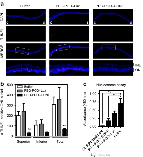
Figure 3.
PEG-POD~GDNF nanoparticles result in decreased apoptosis of photoreceptors. (a) TUNEL stain of retinal sections 48 hours after light exposure showed a significant reduction in apoptotic photoreceptors in PEG-POD~GDNF-injected eyes. C, central; S, superior. (b) TUNEL-positive nuclei in the ONL of both the superior and inferior hemispheres. Eyes injected with PEG-POD~GDNF showed a significant decrease in the number of TUNEL-positive nuclei in the ONL of the superior pole (31.00 ± 8.5 nuclei/section) compared to PEG-POD~Lux (241.2 ± 77.6 nuclei/section, P < 0.01) and buffer- (197.9 ± 39.7, P < 0.01) injected eyes. PEG-POD~GDNF, n = 10; PEG-POD~Lux, n = 5; buffer, n = 7. Mean ± SEM. (c) Retinal tissue was harvested 48 hours post-light treatment and apoptosis evaluated by nucleosome release ELISA. Lower absorbance is observed with less nucleosome release and is thus an indicator of decreased apoptosis. Eyes injected with PEG-POD~GDNF showed 2.3-fold lower absorbance than PEG-POD~Lux-injected eyes (P > 0.05) and 3.9-fold lower than buffer-injected eyes (P < 0.05). Nonlight treated and PEG-POD~GDNF, n = 5; PEG-POD~Lux, buffer, and no light treatment, n = 5. Mean ± SEM. GDNF, glial cell line–derived neurotrophic factor; INL/ONL, inner/outer nuclear layer; PEG, polyethylene glycol; POD, peptide for ocular delivery; TUNEL, TdT-dUTP terminal nick-end labeling.

