Figure 4.
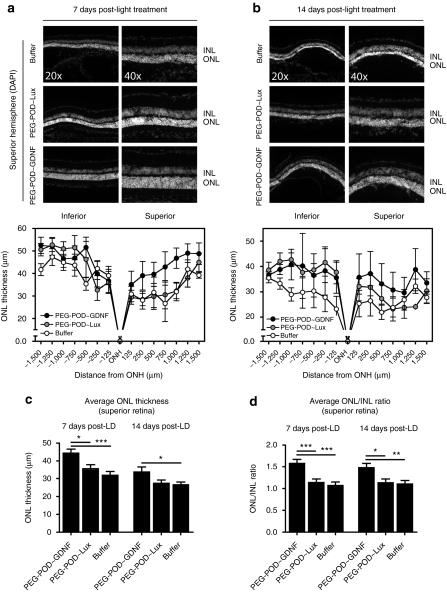
Injection of PEG-POD~GDNF results in decreased photoreceptor cell loss. To examine the effect on photoreceptor cell loss, ONL thickness was measured in retinas injected with either PEG-POD~GDNF, PEG-POD~Lux, or buffer harvested (a) 7 days or (b) 14 days post-light exposure. ONL thickness was measured at 250 µm intervals extending from the optic nerve. Representative images of the superior hemisphere adjacent to the ONH are shown. Average thickness of the ONL of the superior retina was calculated from measurements taken between 250 and 1,500 µm from the optic nerve. Similar measurements were taken for the INL and the average ONL/INL ratio calculated. (c) PEG-POD~GDNF-injected eyes showed a significant increase in the ONL thickness of the superior hemisphere by 24.5% when compared to PEG-POD~Lux (P < 0.05) or by 39.3% buffer (P < 0.001)-injected eyes 7 days post-light treatment and 27.7% relative to buffer (P < 0.05) 14 days post-light treatment. (d) Similar results were obtained when analyzing the ONL/INL ratio of the superior hemisphere. At 7 days the ratio was higher in eyes injected with PEG-POD~GDNF by 38.5% compared to those injected with PEG-POD~Lux (P < 0.001) or 47.3% compared to buffer (P < 0.001). The ONL/INL ratio remained higher at 14 days post-light exposure in PEG-POD~GDNF-treated eyes by 30.4% compared to PEG-POD~Lux (P < 0.05) and by 33.9% compared to buffer (P < 0.01). 7 days: PEG-POD~GDNF and PEG-POD~Lux, n = 6; buffer, n = 4. 14 days: PEG-POD~GDNF, n = 4; PEG-POD~Lux, n = 7; buffer, n = 6. Mean ± SEM. DAPI, 4′,6-diamidino-2-phenylindole; GDNF, glial cell line–derived neurotrophic factor; INL, inner nuclear layer; LD, light degeneration; ONH, optic nerve head; ONL, outer nuclear layer; PEG, polyethylene glycol; POD, peptide for ocular delivery.

