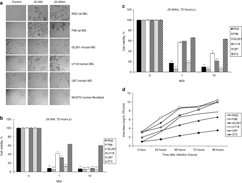
Figure 1.
JX-594 and JX-594m infect and kills human and murine brain tumor cells in vitro. (a) CPE of MGs cells. Cells were plated at confluency and the next day infected with JX-594/JX-594m at an MOI of 10. Microscopy was performed 72 hours after viral infection (original magnification × 100). (b,c) MTT assay of MG cells compared to NIH3T3 controls 72 hours after (b) JX-594 and (c) JX-594m. (d) Viral titers were obtained in MGs and NIH3T3 cell lines after JX-594 infection. Viral titers were determined using a standard plaque titration assay on U2OS cells. Values represent mean PFUs ± SD from triplicate wells. *P < 0.05; **P < 0.01; ***P < 0.001 as analyzed by two-way ANOVA. ANOVA, analysis of variance; CPE, cytopathic effect; MG, malignant glioma; MOI, multiplicity of infection; p.i., postinfection.

