
Fig. 5.
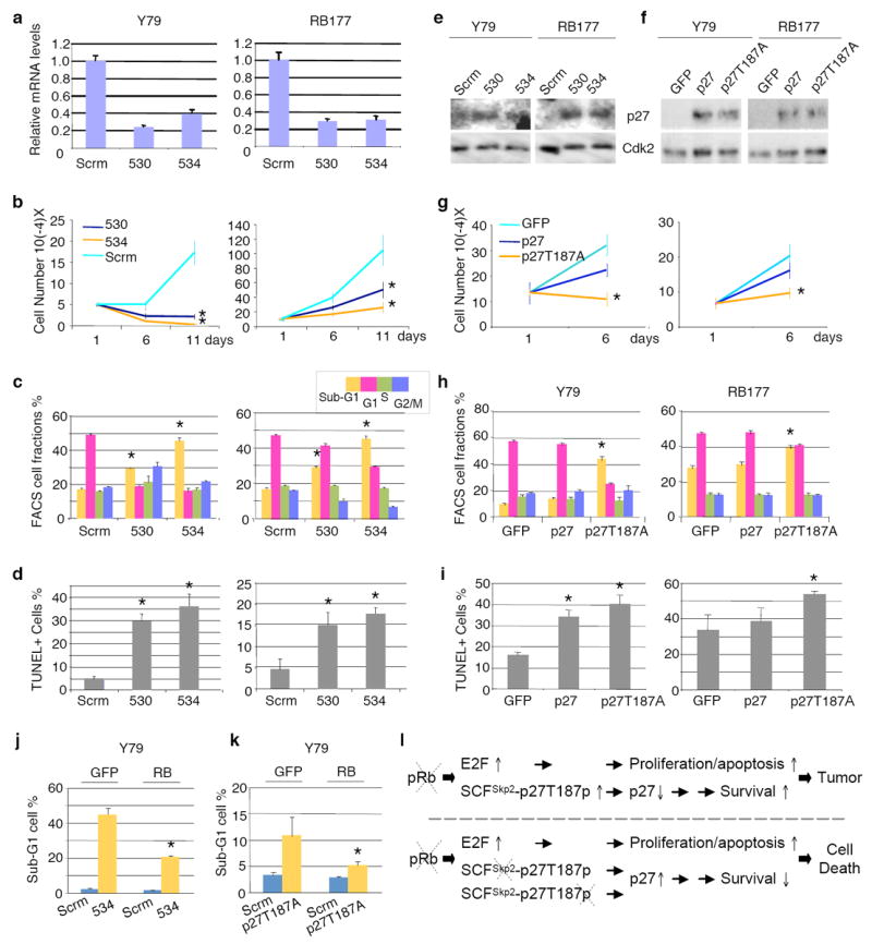
Effects of Skp2 knockdown and stabilized p27 expression on established Y79 cells and early passage RB177 retinoblastoma cells. (a-e) Y79 and RB177 cells infected with lentiviruses expressing shRNA targeting Skp2. Two independent Skp2 shRNAs and a scrambled shRNA control (Scrm) were used as indicated. After drug selection, infected cells were evaluated for Skp2 mRNA by quantitative RT-PCR (a), cell proliferation by counting live cells (b), cell cycle profile by FACS (c), apoptosis by TUNEL staining (d), and p27 expression by Western immunoblotting, with Cdk2 as a loading control (e). (f-i) Y79 and RB177 cells infected with BE-GFP lentiviral vector encoding p27 or p27T187A. Infected cells were evaluated for p27 expression (f), cell proliferation (g), cell cycle profile (h), and TUNEL staining (i), (j-k) Y79 cells transduced with BE-GFP vector or BE-GFP-RB, followed 2 days later by transduction with Skp2 shRNA or scrambled shRNA control (j) or with BE-GFP or BE-GFP-p27T187A (k), and evaluated cells with sub-G1 DNA content. Averages with s.d. are shown. Asterisks indicate P < 0.05 relative to applicable controls. l. A new model of tumorigenesis after Rb1 loss. Two consecutive arrows suggest the presence of multiple steps between them.