Fig. 1.
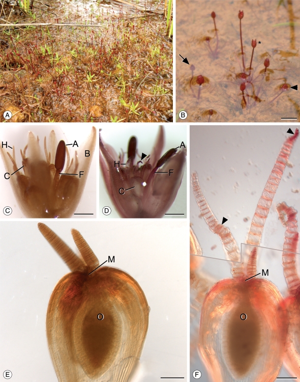
Morphology and development of Trithuria submersa. (A) Habit of T. submersa at Kulunilup Swamp, Kulunilup Nature Reserve. T. submersa plants are red (bright green leaves are immature Goodenia claytoniacea). (B) Reproductive units at various stages of emergence; completely submerged (stage 1; arrow), at the water level creating a depression (stage 2; arrowhead) and fully emergent (stage 3; asterisk). Scale bar = 5 mm. (C) One reproductive unit showing four bracts surrounding carpels with elongating stigmatic hairs and a single central stamen comprising a partially elongated filament and non-dehiscent anther (developmental stage 2). Note that one stigmatic hair is typically longer than the others. Scale bar = 500 µm. (D) One reproductive unit with two dehiscent anthers (developmental stage 4). Some stigmatic hairs have received pollen and cells have collapsed, causing stigmatic hairs to bend (arrowhead). Scale bar = 500 µm. (E) A single carpel from a stage 1 reproductive unit. Stigmatic hairs are short and cell length is much greater along the axis perpendicular to the stigmatic hair. Scale bar = 100 µm. (F) A composite image (two focal planes) showing a single carpel from a mature reproductive unit (stage 4) with two elongated and one short stigmatic hair. Stigmatic cells have elongated and some have collapsed (arrowheads). Grey line indicates the border between images. Scale bar = 100 µm. Abbreviations: A, anther; B, bract; C, carpel; F, filament; H, stigmatic hair; M, carpel mouth; O, ovule.

