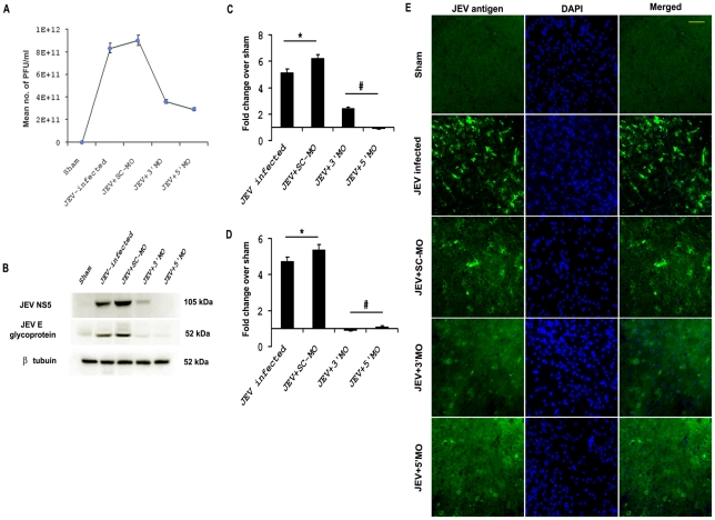
Figure 3. MOs treatment reduces the viral load in vivo.
Significant reduction in viral titer was observed following MO treatment in animals, as compared to JEV-infected and JEV+SC-MO groups. (* p<0.001 for JEV and JEV+SC-MO when compared to Sham, # p<0.001 for JEV+ 3′MO and JEV+ 5′ MO when compared to only JEV-infected group) (A). Immunoblot analysis showed expression of JEV NS5 and E glycoprotein were significantly increased in JEV-infected and JEV+SC-MO than Sham, but reduced significantly after both 3′ and 5′ MO treatments (* p<0.01 for JEV and JEV+SC-MO when compared to Sham, # p<0.01 for JEV+ 3′MO and JEV+ 5′ MO when compared to only JEV-infected group) (B–D). Immunostaining of brain sections from different treatment groups showed greater presence of JEV antigen in JEV and JEV+SC-MO groups (E). Magnification ×20; scale bar correspond to 50µ Photomicrographs shown here in this figure are representative of three individual animals from each group.

