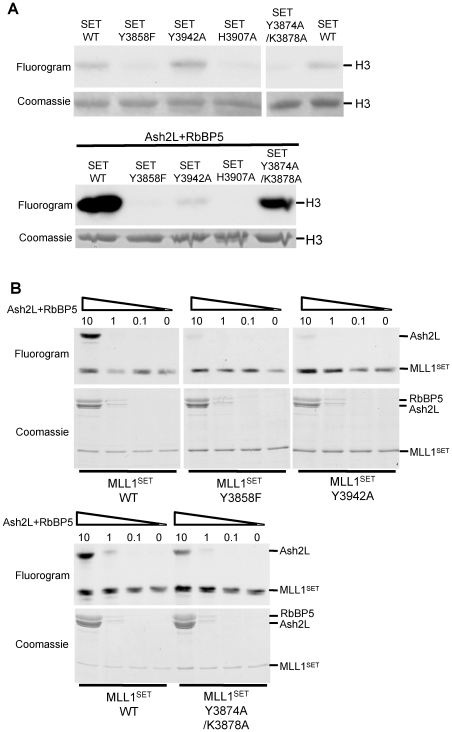
Figure 5. Ash2L/RbBP5 and MLL1SET coordinate in H3 K4 methylation.
(A) In vitro HMT assays using ∼5 µM of wild type and mutants MLL1SET either alone (top) or with Ash2L/RbBP5 (bottom) as enzymes (B) SAM binding assays for Ash2L and either wild type MLL1SET or SET domain mutants. 3 µM of wild type MLL1SET or SET domain mutants was used in each reaction. Molar ratios of Ash2L/RbBP5 versus MLL1SET were indicated on top. The positions for Ash2L and RbBP5 were indicated on left. Duplicate samples were run for Coomassie staining as the loading controls.

