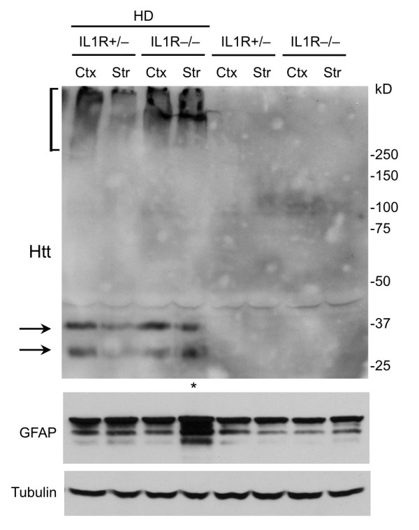
Figure 4.

Western blot analysis of the expression of mutant htt and GFAP in HD mice. Tissue extracts from the cortex (Ctx) and striatum (Str) of HD-IL1R+/- and HD-IL1R-/- mice were subjected to western blotting with EM48. Aggregated htt is presented in the stacking gel (bracket). Soluble mutant htt is indicated by arrows. Note that htt aggregates and soluble mutant htt are more abundant in the HD-IL1R-/- mouse striatum. The same blot was also probed with antibodies to GFAP and tubulin, revealing an increase of GFAP in the HD-IL1R-/- mouse striatum as well.
