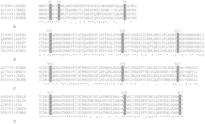
Figure 1. Multiple sequence alignment of amino acid residues at the N and C-termini of PFK1 proteins that form allosteric citrate binding sites.
A. Amino acid residues (grey background) of citrate binding sites in the N-terminal region of PFK1 isoforms from the following species: ECO24, Escherichia coli; ASPNG Aspergillus niger; CAEEL Caenorhabditis elegans; DROME, Drosophila melanogaster; HUMAN, Homo sapiens. B. Amino acid residues (grey background) of citrate binding sites in the C-terminal region of PFK1 isoforms from the following fungi: ASPNG, Aspergillus niger; ASPFU, Aspergillus flavus; Yeast, Saccharomyces cerevisiae; PICPA, Pichia pastoris; SCHPO, Schizosaccharomyces pombe. C. Amino acid residues (grey background) of citrate binding sites in the C-terminal region of PFK1 isoforms from the following invertebrates: SCHMA, Schistosoma mansoni; HAECO, Haemonchus contortus; CAEEL, Caenorhabditis elegans; DROME, Drosophila melanogaster. D. Amino acid residues (grey background) of citrate binding sites in the C-terminal region of PFK1 isoforms from the following species: XENLA, Xenopus laevis; CHICK, Gallus gallus; PIG Sus scrofa; CANFA, Canis familiaris; HUMAN, (Homo sapiens). The components of allosteric citrate site were originally identified in the mouse PFK-C enzyme (Accession number Q9WUA3) [6], which has 69,58% of identical; 13,18% strongly similar and 6,46% weakly similar residues to the human PFK-M (Accession number P08237); however, there is a minor shift in numbering of amino acid residues between the enzymes. The mouse PFK-C enzyme has an extension of 8 amino acid residues at the N-terminal end of the enzyme and an insertion at position 349. Therefore, the corresponding ligand binding sites in the N-terminal part of human PFK-M differ by 8 amino acid residues and in the C-terminal region by 9 residues with respect to the mouse PFK-C. The numbering system for amino acids used in the entire paper, therefore reflects the positions on the human PFK-M. The alignments were generated using CLUSTAL W [34].

