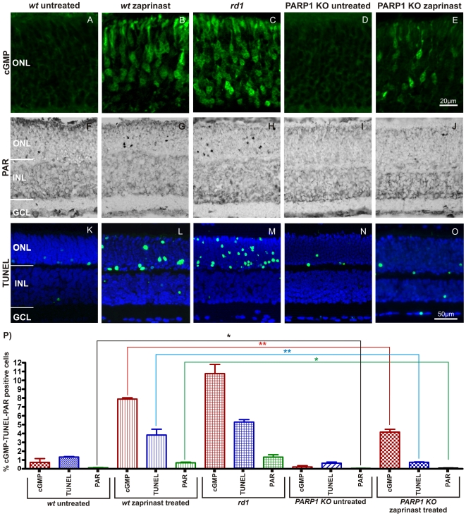
Figure 3. PARP1 KO animals are resistant to PDE6 inhibition induced photoreceptor cell death.
Organotypic retinal explant cultures obtained from wt and PARP1 KO animals were treated with the PDE6 inhibitor zaprinast and compared to untreated wt, rd1 and PARP1 KO cultured retinae. Control wt retinae at P11 in vitro showed minimal immunoreactivity to cGMP-antibody in the ONL (A). Zaprinast treated wt retinae exhibited strongly increased intracellular cGMP levels (B), similar to what was observed in PDE6 mutant rd1 retina (C). While PARP1 KO did not display cGMP immunoreactivity (D), it responded to zaprinast treatment with a moderate, but significant, elevation of cGMP positive ONL cells (E). Accumulation of PAR as an indirect marker for PARP activity (F–J) was found in large amounts only in zaprinast treated wt or rd1 ONL but notably absent in PARP1 KO preparations and wt retinae. The TUNEL assay for dying cells generally followed a similar pattern (K–O): The rd1 mutant or PDE6 inhibition resulted in marked increases of positive cells in the ONL only. The bar graph (P) illustrates the quantification of the three parameters assayed. Explant cultures from n = 3–6 animals were used for each treatment situation and genotype. Error bars represent SEM.

