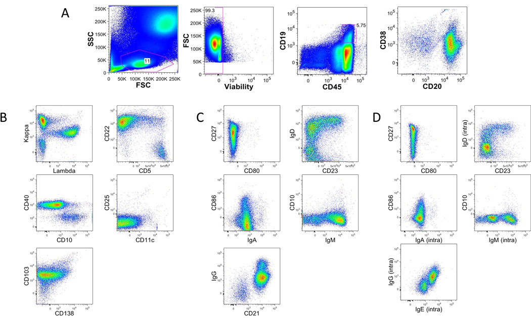
Figure 3. Gating strategy in B lineage tubes.
A. Single-cell suspensions from healthy donors were stained with a combination of 14 antibodies and viability dye as described in the material and methods and table 1 (B1, B2 and B3). Lymphocytes were identified based on their forward and side scatter properties. Subsequently, dead cells were excluded through the use of a viability dye. CD45 and CD19 were used to identify B cells (CD45+CD19+) among the previously selected living lymphocytes.
B. Representative dot plot of the markers specific of Staining B1.
C. Representative dot plot of the markers specific of Staining B2.
D. Representative dot plot of the markers specific of Staining B3.

