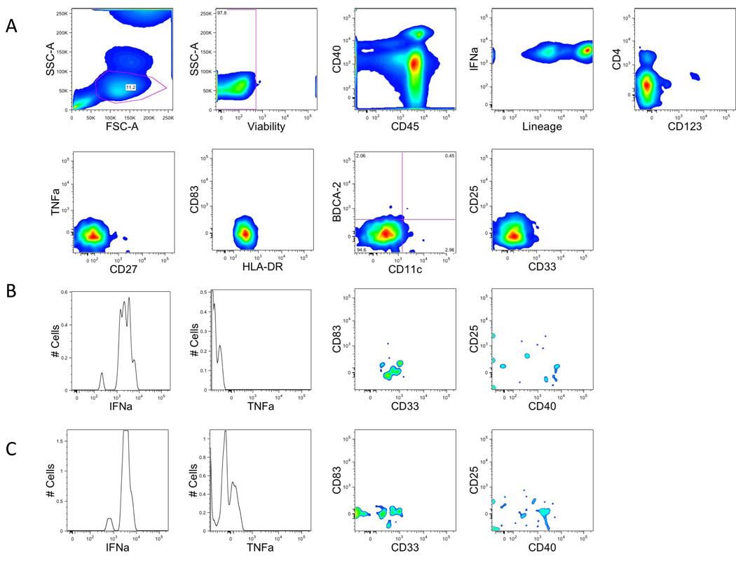
Figure 6. Gating strategy in dendritic cell lineage tubes.
A. Single-cell suspensions from healthy donors were stained with a combination of 14 antibodies and viability dye as described in the material and methods and Table 1. Dendritic cells were identified based on their forward and side scatter properties (same as lymphocyte). Subsequently, dead cells were excluded through the use of a viability dye. CD45 and lineage were used to identify DC cells (CD45−Lineage−). Expression of HLA-DR was required, and we identified myeloid dendritic cells (CD11c+) from plasmacytoid dendritic cells (CD123+) with the expression of CD11c and CD123.
B. Myeloid dendritic cells (CD11c+) were analyzed for the expression of cytokines such as IFNa, TNFa or activation markers CD83, CD25, CD40, CD33
C. Plasmacytoid dendritic cells (CD123+) were analyzed for the expression of cytokines such as IFNa, TNFa or activation markers CD83, CD25, CD40.
Due to low number of cells, all figures are smoothed histograms.

