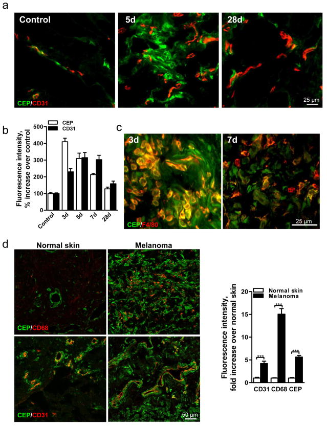
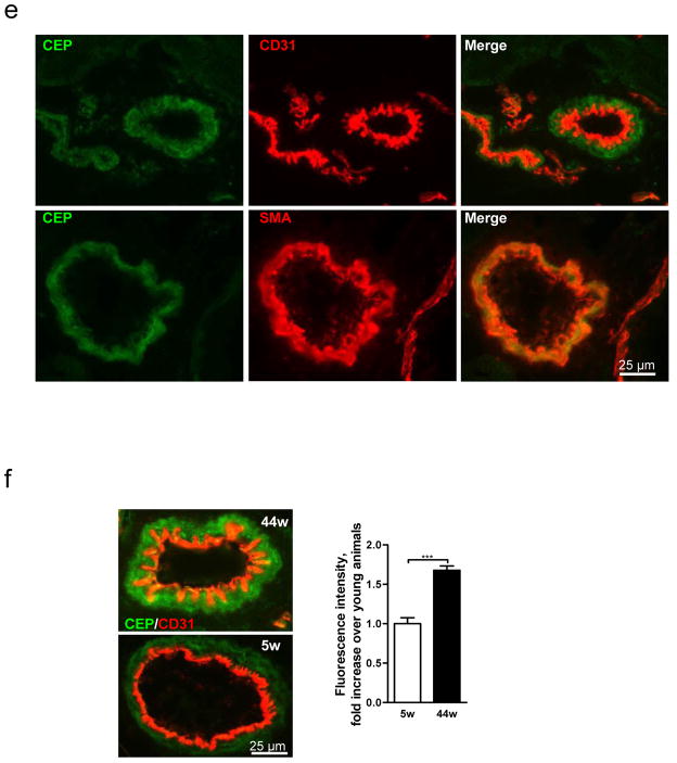
Fig. 1. CEP, an end product of lipid oxidation, is present in wounds, elevated in melanoma and accumulated in aging tissues.
a. CEP and CD31 co-staining in normal and wounded skin 5 and 28 days post-injury. b. Quantified levels of CEP and CD31, n=5. c. F4/80 macrophage marker and CEP distribution in wound tissues 3 and 7 days post-injury. d. CEP and CD68 (top) or CEP and CD31 (bottom) presence in human skin and melanoma. Right- quantified levels of CD31, CD68 and CEP, n=8. e. CEP and CD31 (top) or CEP and SMA (bottom) distribution in murine skeletal muscle. f. CEP and CD31 co-staining in Vastus intermedius sections from 5 and 44 week old mice. Right- CEP quantification, n=4. All values represent mean ± s.e.m. *** p<0.001.


