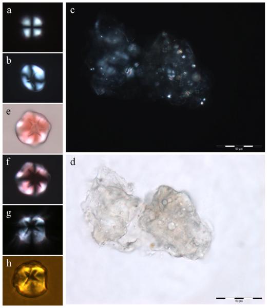
Fig. 1.
Spherulites of Aβ42 formed under near-physiological conditions in vitro. a, spherulite 10-12 μm in diameter with no core; b, spherulite 15-20 μm in diameter with clear core; c, large group of spherulites occluded within an amorphous matrix of Aβ42; d, the same group viewed by optical microscopy; e, spherulite 15-20 μm in diameter stained with Congo red; f, the same spherulite under the polarising microscope; g, spherulite 15-20 μm in diameter stained with ThT viewed by optical microscopy and crossed polarisers; h, the same spherulite stained with ThT viewed by fluorescence microscopy.

